Anatomy
The kidney and stone formation
A stone forms in the urine-collecting region of the kidney (the renal pelvis). When it passes from here into the ureter, it blocks urine flow and causes severe pain.
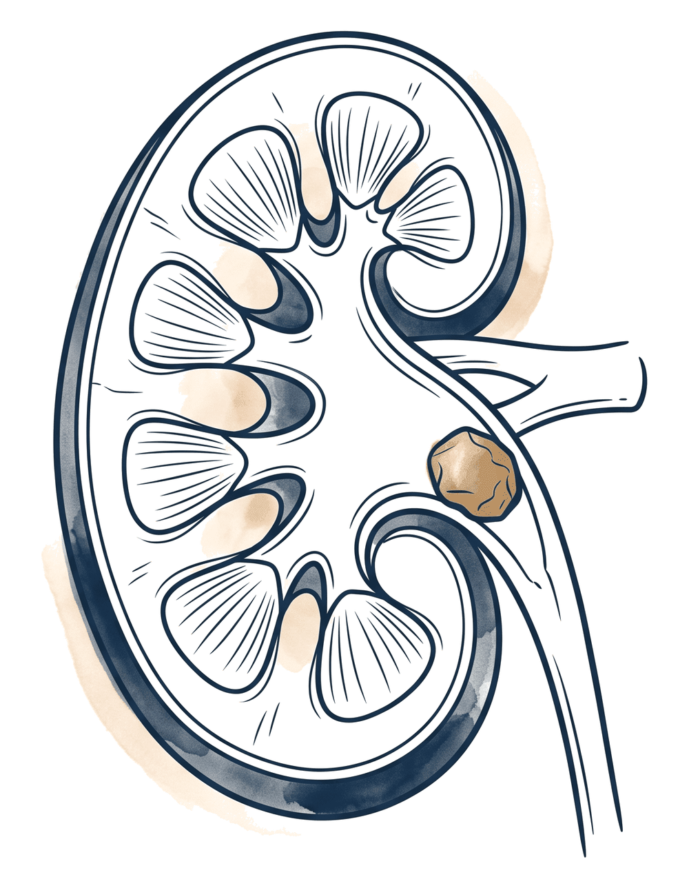
One of the most common causes of severe flank pain. Today, most kidney stones can be treated effectively with incision-free, minimally invasive methods.
A kidney stone forms when minerals and salts in the urine crystallize and accumulate into hard masses. Stones can be as small as a grain of sand, or grow into large "staghorn" stones that fill the kidney's collecting system.
Small stones often pass on their own with the urine. But when a stone lodges in the urinary tract (the ureter), it blocks urine flow and causes severe pain (renal colic). This pain is described as one of the most intense known.
Kidney stones are quite common in the population, and people who have passed a stone once are at high risk of recurrence. For this reason, not only treating the stone but also investigating the cause to prevent recurrence is important.
A stone forms in the urine-collecting region of the kidney (the renal pelvis). When it passes from here into the ureter, it blocks urine flow and causes severe pain.
Symptoms vary with the size and location of the stone. While some stones stay silent, obstructing stones produce a typical picture.
Excruciating pain radiating from the flank to the side and groin, coming in waves (renal colic).
Pink, red or tea-colored urine; caused by the stone irritating the urinary tract.
Severe pain is often accompanied by nausea and vomiting.
As the stone approaches the bladder, a frequent, small-volume and burning urge to urinate may appear.
Fever appears when infection is added — this is an emergency and must be treated immediately.
Cloudiness and a bad smell in the urine may point to an accompanying infection.
Diagnosing a kidney stone is fast and clear. The right imaging also determines the treatment plan.
The type and radiation of the pain are typical of a stone. Blood and infection are assessed with a urine test.
Non-contrast computed tomography is the most definitive method showing the location and size of the stone.
A radiation-free option; it can show swelling in the kidney (hydronephrosis) and the stone.
In recurrent stones, blood and urine analyses are done to find the cause and take preventive measures.
The most common questions about pain, passing a stone and preventing recurrence.
No. Generally, most stones smaller than 5 mm pass on their own with plenty of fluids and, when needed, medication support. Surgery or lithotripsy comes into play if the stone is large, causes obstruction, does not pass, or there is infection.
ESWL is applied from outside the body and is usually done under sedation. In a suitable patient it is safe and effective. Not every stone is suitable for ESWL; the location, size and hardness of the stone are important in deciding the method.
No. In RIRS and URS methods, entry is through the urinary tract and no skin incision is made. The stone is turned to dust with the laser. Recovery is fast, and most patients return to daily life in a short time.
The most effective measure is adequate fluid intake — aim for 2–3 liters of water a day. Reducing salt, eating a balanced diet and, in recurrent stones, analyzing the stone's content to take cause-specific measures are important.
If severe pain is accompanied by fever and chills, if you cannot pass urine, or if you have a single kidney, seek help without delay. A stone obstructing with infection is an emergency.
With the right imaging, the size and location of the stone become clear; let's plan together the most suitable, least invasive treatment for you. You can reach out via WhatsApp for a preliminary consultation.